Quest Online Homework due 5/10 and 5/17
Resistance of a Light Bulb lab due 5/19
Unit Test 5/20
Current ElectricityUnit Goals
Know meanings of potential difference (voltage), current, resistance, power. Be able to use appropriate relationships between them with correct abbreviations and units.
Properly use the terms series, parallel and circuit.
Draw and decipher circuit diagrams.
Determine current, resistance, potential difference and power output for any part of a circuit, including
Simple circuits, series circuits, parallel circuits and complex circuits with both series and parallel elements.
Describe what affects an object’s resistance and categorize resistors as ohmic or nonohmic
Interpret graphs about resistance I vs. V and V vs. R.
Know how to include ammeters and voltmeters in a circuit and what this says about their resistances.
Describe the structure of a capacitor.
Use transient currents to describe how steady state voltages and currents are established.
Describe how houses are wired and the role of circuit breakers.
Determine loss of energy to heat in wires and describe how it can be reduced.
Trace the conducting path through light bulbs.
Describe the production of electrical energy in batteries and the role of internal resistance.
Use the pressure metaphor for voltage and interpret color-coded voltage diagrams.
Kamis, 28 April 2011
Calibrating a pH meter using buffers
A pH meter requires proper calibration in order to give accurate pH readings. The meter must accuratetely translate voltage measurements into pH measurements.
How a pH meter is calibrated to give accurate pH readings?
Procedure for an 2-point calibration:
The third point is used to confirm that the pH meter has been calibrated correctly.
8F6XDHBGS4VN
How a pH meter is calibrated to give accurate pH readings?
A pH meter is calibrated by immersing its electrode(s) into buffers (test solutions of known pH) and by adjusting the meter accordingly. Since pH measurements are affected by temperature, the temperature must remain constant during the time of the calibration. Modern pH meters have build-in thermometers and automatically correct their own pH measurements as the temperature changes.
See the following video to get an idea how a pH meter is calibrated using buffer solutions:
What are the main steps for a generic pH meter calibration?
The following procedures are used to calibrate pH meters:
· A 2 or 3-point calibration : Two or three buffers solutions are used respectively. They are usually sufficient for initial calibration. After this initial calibration the meter can accurately measure pH values in between.
· A 1-point calibration: A buffer with a pH close to the expected sample pH is used. The pH measurements are not as accurate as with the 2 or 3 point calibration.
Procedure for an 1-point calibration:
- Place the pH buffer solution (normally pH = 4.01 solution) into a small beaker. Place a magnetic stirrer and a temperature probe into the buffer solution in the beaker.
- Measure the temperature of the buffer solution if the pH meter does not have its own temperature probe. Adjust the temperature control of the pH meter according to the buffer’s measured temperature (remember pH measurements are temperature dependent).
- Remove the electrode protective cap(s). Rinse the electrodes and the temperature probe with distilled water using a squeeze bottle.
- Dab dry the bottom of the glass bulb with a tissue paper. Do not wipe the glass bulb (scratches and static charges affect the electrode’s response).
- Place the electrode(s) and temperature probe into the well stirred pH buffer solution. The porous frit must be covered with the buffer solution.
- Adjust the slope/sensitivity control to read the true pH of the buffer solution (modern pH meters do this automatically).
Procedure for an 2-point calibration:
- Place the pH buffer solution (normally pH = 7.0 solution) into a small beaker. Place a magnetic stirrer and a temperature probe into the buffer solution in the beaker.
- Follow steps 2-6 of the 1-point calibration.
- Follow steps 1-6 of the 1-point calibration using the pH = 4.01 buffer solution
- Keep repeating steps 2 and 3 until practically no adjustments are required
Procedure for a 3-point calibration:
- Follow steps 1-6 of the 2-point calibration procedure
- Use a third pH buffer, whose pH value is as close as possible to the suspected pH of the sample, and follow the 1-point calibration.
8F6XDHBGS4VN
Rabu, 27 April 2011
Stanville Primary School - 27th April 2011 - KESH Academy Physics Factory
8 Year Five Stanville Students are attending a five-week Physics course at the KESH academy Physics Factory. They have studied sound, magnets and rocketry!
Selasa, 26 April 2011
Measuring the pH of a Solution with a pH meter
The pH of a solution can be measured quickly and accurately with a pH meter (see Figure 1).
How does a pH meter work?
The main limitations of the glass electrode are:
 |
| Figure 1: A digital pH meter |
How does a pH meter work?
A pH meter has to somehow measure the concentration of the hydrogen ions [H+] in a solution. An acidic solution has far more positively charged hydrogen ions in it than an alkaline solution, so it has greater potential to produce an electric current under certain conditions - in other words, it is like a battery that can produce a greater voltage. A pH meter takes advantage of this and works like a typical voltameter: in brief, a pH meter consists of a pair of electrodes connected to a meter capable of measuring small voltages, on the order of millivolts. It measures the voltage (electrical potential) produced by the solution whose acidity we are interested in, compares it with the voltage of a known standard solution, and uses the difference in voltage (the potential difference) between them to calculate the difference in pH.
What are the parts of a pH meter?
A typical pH meter consists of two parts: i) one special measuring probe (a glass electrode) or two measuring probes that are inserted into the solution whose pH is required and ii) an electronic meter that measures and displays the pH reading.
A glass electrode is in a sense two electrodes combined in one. It consists of a long glass tube with a thin walled glass bulb at the end. Special glass of high electrical conductance and low melting point is used for the purpose. This glass can specifically sense hydrogen ions H+ up to a pH ≈ 9 (with special glass electrodes pH ranges from 1-13 can be measured). The bulb contains 0.1 M HCl and a Ag/AgCl electrode (used as an internal reference electrode) is immersed into the solution and connected by a platinum wire for electrical conduct.
 |
| Figure 2: A glass electrode |
The main advantages of the glass electrode are:
- It can be used in the presence of strong oxidizing or reducing substances and metal ions
- Accurate results are obtained in the range pH 1-9. However, by using special glass electrodes pH 1-13 can be measured
- It is simple to operated. It can be attached to portable instruments and is used quite often in chemical, biological, industrial and agricultural laboratories
- It does not function properly in some organic solvents (i.e. ethanol)
- It does not function properly above pH > 9 since it is sensitive to Na+ ions so a correction has to be made
In case that the pH meter has two probes (two electrodes): i) one of them is a glass electrode (has silver wire suspended in a solution of KCl that is contained in a special glass bulb coated with silica and metal salts) and ii) the other is the reference electrode and has a KCl wire suspended in a solution of KCl (see Figure 3).
When the probe(s) are immersed into the solution some of the H+ ions in the solution move toward the glass electrode (Figure 3, labeled as 2) and replace some of the metal ions in its special surface. This creates a tiny current (voltage) that the silver electrode passes to the measuring device, the voltameter. The voltameter measures the voltage generated and shows a corresponding pH measurement as follows:
· A higher voltage means more H+ ions in the solution and therefore a higher acidity. The pH meter shows in such a case a lower pH value since the solution is more acidic.
· A lower voltage means fewer H+ ions in the solution and therefore a lower acidity. The pH meter shows in such a case a higher pH value since the solution is less acidic or better more alkaline.
The reference electrode (Figure 3, labeled as 6) acts as a reference for the measurement.
How accurate measurements can be made with a pH meter?
Saprophytic Orchids of Indiana
There are four species of orchids with extant populations in Indiana that can be considered to be saprophytic and/or hemi-parasitic. They live their lives below ground, not subject to photosynthesis, and unable to produce their own food, but deriving it instead (via mycorrhizae) from decaying organic matter (Homoya 1993).
It is suggested that no plants are truly saprophytic, that it is not the plants that are doing the breaking down of the dead plant material, but symbiotic fungi (mycorrhiza) working with the plants that are doing the decomposing (www.helium.com).
I was fortunate to find and photograph all four species last year, but it required traveling from one end of the state to the other, beginning the first week in May and ending in the middle of September.
The first to bloom in the spring is Wister's coral-root (Corallorhiza wisteriana), which is found primarily in the southern half of the state. The flowers (below) were found in bloom on May 4, near Versailles State Park and were growing in the flood plain of a small creek and not on the moderately moist slopes of ravines where it typically occurs (Homoya 1993).


Later in the summer in late July and August one can find spotted coral-root (Corallorhiza maculata). The flowers (below) were blooming on July 18, at Cowles Bog in northwestern Indiana.


Another mid- to late-summer bloomer is crested coral-root (Hexalectris spicata), which differs from members of the coral-root genus (Corallorhiza) by having a different column structure and thicker more unbranched rhizomes (Homoya 1993). The flowers (below) were photographed on July 20, in Clark County.


Autumn coral-root (Corallorhiza odontorhiza) is the smallest member of this genus and no doubt possesses the least showy flower, if indeed the flower can even be found in its open state. I have been monitoring a small colony of this species at Cowles Bog in Porter County for four years. Only once have I found a single open flower on any of the plants.
This is the typical flower stalk of autumn coral-root (below) with its tightly closed flower.


And here--in all its glory--is the open flowered form (below) photographed on September 19.

Sadly, another saprophytic orchid, early spring coral-root (Corallorhiza trifida), has been extirpated from Indiana. It was found in one site only, in a unique natural area in the Indiana Dunes, which is now the location of a foreign owned steel mill (Homoya 1993).
Homoya, M.A. 1993. Orchids of Indiana. Indianapolis: The Indiana Academy of Science.
It is suggested that no plants are truly saprophytic, that it is not the plants that are doing the breaking down of the dead plant material, but symbiotic fungi (mycorrhiza) working with the plants that are doing the decomposing (www.helium.com).
I was fortunate to find and photograph all four species last year, but it required traveling from one end of the state to the other, beginning the first week in May and ending in the middle of September.
The first to bloom in the spring is Wister's coral-root (Corallorhiza wisteriana), which is found primarily in the southern half of the state. The flowers (below) were found in bloom on May 4, near Versailles State Park and were growing in the flood plain of a small creek and not on the moderately moist slopes of ravines where it typically occurs (Homoya 1993).


Later in the summer in late July and August one can find spotted coral-root (Corallorhiza maculata). The flowers (below) were blooming on July 18, at Cowles Bog in northwestern Indiana.


Another mid- to late-summer bloomer is crested coral-root (Hexalectris spicata), which differs from members of the coral-root genus (Corallorhiza) by having a different column structure and thicker more unbranched rhizomes (Homoya 1993). The flowers (below) were photographed on July 20, in Clark County.


Autumn coral-root (Corallorhiza odontorhiza) is the smallest member of this genus and no doubt possesses the least showy flower, if indeed the flower can even be found in its open state. I have been monitoring a small colony of this species at Cowles Bog in Porter County for four years. Only once have I found a single open flower on any of the plants.
This is the typical flower stalk of autumn coral-root (below) with its tightly closed flower.


And here--in all its glory--is the open flowered form (below) photographed on September 19.

Sadly, another saprophytic orchid, early spring coral-root (Corallorhiza trifida), has been extirpated from Indiana. It was found in one site only, in a unique natural area in the Indiana Dunes, which is now the location of a foreign owned steel mill (Homoya 1993).
Homoya, M.A. 1993. Orchids of Indiana. Indianapolis: The Indiana Academy of Science.
Senin, 25 April 2011
Measuring the pH of a solution / Acid-Base Indicators
The pH of a solution can be measured as follows:
What kind of substances are acid-base indicators?
Acid-base indicators are usually weak organic acids or weak organic bases. They tend to have different color depending on the pH of the solution in which they are in.
How acid-base indicators are prepared in the lab?
These are usually solid substances that are dissoved in a solvent (i.e. ethanol). Few drops of the solution of the indicator is added to the solution that we would like to determine the pH.
How simple acid-base indicators work?
In reality what happens is that the two forms of the indicator participate in an equilibrium:
ΗMeo + H2O Meo- + H3O+ [1]
where ka is the ionization constant of methyl orange.
As a conclusion for monoprotic indicators of the general structure ΗIn with an equilibrium constant ka:
- By Acid-Base Indicators (less precise)
- By a pH-meter
What kind of substances are acid-base indicators?
Acid-base indicators are usually weak organic acids or weak organic bases. They tend to have different color depending on the pH of the solution in which they are in.
How acid-base indicators are prepared in the lab?
These are usually solid substances that are dissoved in a solvent (i.e. ethanol). Few drops of the solution of the indicator is added to the solution that we would like to determine the pH.
How simple acid-base indicators work?
An acid - base indicator is a colored substance that itself can exist in either an acid or base form. The acid form has a different color than the base form. Thus, the indicator turns one color in an acidic solution and another color if placed in a basic solution. If you know the pH at which the indicator turns from one form to the other, you can determine whether a solution has a higher or lower pH than this value.
For example methyl orange is one of the indicators commonly used in titrations. It gradually changes color from red to yellow over the pH interval from 3.1-4.4. In a solution with a pH > 4.4 exists as a species with negative charge (anion, Meo- ) and has a yellow color. In a solution with a pH < 3.1 exists in its neutral form and haw a red color (ΗMeo).
In reality what happens is that the two forms of the indicator participate in an equilibrium:
ΗMeo + H2O
If acid is added the position of the above equilibrium shifts to the left according to Le Chatelier's Principle and turns the indicator red (the solution takes a red color).
If base is added the position of the equilibrium shifts to the right according to Le Chatelier's Principle and turns the indicator yellow (the solution takes a yellow color).
The Ηenderson-Hasselbach equation can be used in order to determine the pH range an indicator changes color. Let's apply this for the methyl orange case:
pH = pka + log [Meo-] / [ΗMeo] [2]
It has been determined experimentally that when 90% or more of the indicator is in the ΗMeo form (that means when the ratio [Meo-] / [ΗMeo] ≈ 0,1) then the color of the solution is red. If 90% or more of the indicator is in the Meo- form (that means [Meo-] / [ΗMeo] ≈ 10) the the color of the solution becomes yellow. By subsituting the above ratios to the Ηenderson-Hasselbach equation the pH range an indicator changes color can be determined:
pH = pka + log [Meo-] / [ΗMeo] = pka + log(0,1) = pka – 1 [3]
και pH = pka + [Meo-] / [ΗMeo] = pka + log(10) = pka + 1 [4]
When [Meo-] = [ΗMeo] the color of the indicator is a mixture of yellow and red and the solution takes an orange color.
From equation [3] and [4 ] it can be determined that the indicator changes color over a range of two pH units (when the pH is between pka + 1 and pka - 1).
If pH < pka – 1, then the color of the solution takes the color of the ΗI form (unionized form)
If pH > pka + 1, then the color of the solution takes the color of the I- (ionized form)
If pH = pka then the color of the solution is a «mixture» of the colors of ΗI and I-.
In the video below the color change of an indicator is shown as the pH of the solution in which is in changes from neutral (pH of distilled water) to basic and then back from basic to acidic:
Weird Facts
Can you feel the pulse in your wrist? For humans the normal pulse is 70 heartbeats per minute. Elephants have a slower pulse of 27 and for a canary it is 1000!
If all the blood vessels in your body were laid end to end, they would reach about 60,000 miles.
Abraham Lincoln probably had a medical condition called Marfans syndrome. Some of its symptoms are extremely long bones, curved spine, an arm span that is longer than the persons height, eye problems, heart problems and very little fat. It is a rare, inherited condition.
In one day your heart beats 100,000 times.
By the time you are 70 you will have easily drunk over 12,000 gallons of water.
Coughing can cause air to move through your windpipe faster than the speed of sound - over a thousand feet per second!
Germs only cause disease, right? But a common bacterium, E. coli, found in the intestine helps us digest green vegetables and beans (also making gases - pew!). These same bacteria also make vitamin K, which causes blood to clot. If we didn't have these germs we would bleed to death whenever we got a small cut!
It takes more muscles to frown than it does to smile.
That dust on rugs and your furniture is not only dirt. It's mostly made of dead skin cells. Everybody loses millions of skin cells every day which fall on the floor and get kicked up to land on all the surfaces in a room. You could say, "That's me all over."
It takes food seven seconds to go from the mouth to the stomach via the oesophagus.
A human's small intestine is 6 meters long.
The human body is 75% water.
Your blood takes a very long trip through your body. If you could stretch out all of a human's blood vessels, they would be about 60,000 miles long. That's enough to go around the world twice.
The width of your armspan stretched out is the length of your whole body.
The average human dream lasts only 2 to 3 seconds.
The average American over fifty will have spent 5 years waiting in lines.
The farthest you can see with the naked eye is 2.4 million light years away! (140,000,000,000,000,000,000 miles.) That's the distance to the giant Andromeda Galaxy. You can see it easily as a dim, large gray "cloud" almost directly overhead in a clear night sky.
The average person has at least seven dreams a night.
Your brain is move active and thinks more at night than during the day.
Your brain is 80% water.
85% of the population can curl their tongue into a tube.
Your tongue has 3,000 taste buds.
Your forearm (from inside of elbow to inside of wrist) is the same length as your foot.
A sneeze travels at over 100 miles per hour. Gesundheit!
Your thigh bone is stronger than concrete.
Your fingernails grow almost four times as fast as your toenails.
You blink your eyes over 10,000,000 a year.
There were about 300 bones in your body when you were born, but by the time you reach adulthood you only have 206.
There were about 300 bones in your body when you were born, but by the time you reach adulthood you only have 206.
Exercise Electrical Energy and Power
Minggu, 24 April 2011
Nucleic Acids
If the primary structure of polypeptides determines the conformation of a protein, what determines primary structure? The amino acid sequence of a polypeptide is programmed by a unit of inheritance known as a gene. Genes consist of DNA, which is a polymer belonging to the class of compounds known as nucleic acids.
The Roles of Nucleic Acids
There are two types of nucleic acids: deoxyribonucleic acid (DNA) and ribonucleic acid (RNA) . These are the molecules that enable living organisms to reproduce their complex components from one generation to the next. Unique among molecules, DNA provides directions for its own replication. DNA also directs RNA synthesis and, through RNA, controls protein synthesis.
The figure above shows DNA → RNA → protein: a diagrammatic overview of information flow in a cell. In a eukaryotic cell, DNA in the nucleus programs protein production in the cytoplasm by dictating the synthesis of messenger RNA (mRNA), which travels to the cytoplasm and binds to ribosomes. As a ribosome (greatly enlarged in this drawing) moves along the mRNA, the genetic message is translated into a polypeptide of specific amino acid sequence.
DNA is the genetic material that organisms inherit from their parents. Each chromosome contains one long DNA molecule, usually consisting of from several hundred to more than a thousand genes. When a cell reproduces itself by dividing, its DNA molecules are copied and passed along from one generation of cells to the next. Encoded in the structure of DNA is the information that programs all the cell’s activities. The DNA, however, is not directly involved in running the operations of the cell, any more than computer software by itself can print a bank statement or read the bar code on a box of cereal. Just as a printer is needed to print out a statement and a scanner is needed to read a bar code, proteins are required to implement genetic programs. The molecular hardware of the cell—the tools for most biological functions—consists of proteins. For example, the oxygen carrier in the blood is the protein haemoglobin, not the DNA that specifies its structure.
How does RNA, the other type of nucleic acid, fit into the flow of genetic information from DNA to proteins? Each gene along the length of a DNA molecule directs the synthesis of a type of RNA called messenger RNA (mRNA). The mRNA molecule then interacts with the cell’s protein–synthesizsng machinery to direct the production of a polypeptide. We can summarise the flow of genetic information as DNA → RNA → protein. The actual sites of protein synthesis are cellular structures called ribosomes. In a eukaryotic cell, ribosomes are located in the cytoplasm, but DNA resides in the nucleus. Messenger RNA conveys the genetic instructions for building proteins from the nucleus to the cytoplasm. Prokaryotic cells lack nuclei, but they still use RNA to send a message from the DNA to the ribosomes and other equipment of the cell that translate the coded information into amino acid sequences.
The Structure of Nucleic Acids
Nucleic acids are macromolecules that exist as polymers called polynucleotides.The components of nucleic acids.
(a) A polynucleotide has a regular sugar–phosphate backbone with variable appendages, the four kinds of nitrogenous bases. RNA usually exists in the form of a single polynucleotide, like the one shown here. (
b) A nucleotide monomer is made up of three components: a nitrogenous base, a sugar, and a phosphate group, linked together as shown here. Without the phosphate group, the resulting structure is called a nucleoside.
(c) The components of the nucleoside include a nitrogenous base (either a purine or a pyrimidine) and a pentose sugar (either deoxyribose or ribose).
As indicated by the name, each polynucleotide consists of monomers called nucleotides . A nucleotide is itself composed of three parts: a nitrogenous base, a pentose (five–carbon sugar), and a phosphate group. The portion of this unit without the phosphate group is called a nucleoside.
The DNA double helix and its replication. The DNA molecule is usually double–stranded, with the sugar–phosphate backbone of the antiparallel polynucleotide strands (symbolized here by blue ribbons) on the outside of the helix. Holding the two strands together are pairs of nitrogenous bases attached to each other by hydrogen bonds. As illustrated here with symbolic shapes for the bases, adenine (A) can pair only with thymine (T), and guanine (G) can pair only with cytosine (C). When a cell prepares to divide, the two strands of the double helix separate, and each serves as a template for the precise ordering of nucleotides into new complementary strands (orange). Each DNA strand in this figure is the structural equivalent of the polynucleotide diagrammed below.
DNA double helix
The RNA molecules of cells consist of a single polynucleotide chain like the one shown in the figure above .In contrast, cellular DNA molecules have two polynucleotides that spiral around an imaginary axis, forming a double helix.
The figure above shows the DNA double helix and its replication. The DNA molecule is usually double–stranded, with the sugar–phosphate backbone of the antiparallel polynucleotide strands (symbolised here by blue ribbons) on the outside of the helix. Holding the two strands together are pairs of nitrogenous bases attached to each other by hydrogen bonds. As illustrated here with symbolic shapes for the bases, adenine (A) can pair only with thymine (T), and guanine (G) can pair only with cytosine (C). When a cell prepares to divide, the two strands of the double helix separate, and each serves as a template for the precise ordering of nucleotides into new complementary strands (orange). Each DNA strand in this figure is the structural equivalent of the polynucleotide in the diagram.
James Watson and Francis Crick, working at Cambridge University, first proposed the double helix as the three–dimensional structure of DNA in 1953. The two sugar–phosphate backbones run in opposite 5′ → 3′ directions from each other, an arrangement referred to as antiparallel, somewhat like a divided highway. The sugar–phosphate backbones are on the outside of the helix, and the nitrogenous bases are paired in the interior of the helix. The two polynucleotides, or strands, as they are called, are held together by hydrogen bonds between the paired bases and by van der Waals interactions between the stacked bases. Most DNA molecules are very long, with thousands or even millions of base pairs connecting the two chains. One long DNA double helix includes many genes, each one a particular segment of the molecule.
Only certain bases in the double helix are compatible with each other. Adenine (A) always pairs with thymine (T), and guanine (G) always pairs with cytosine (C). If we were to read the sequence of bases along one strand as we traveled the length of the double helix, we would know the sequence of bases along the other strand. If a stretch of one strand has the base sequence 5′–AGGTCCG–3′, then the base–pairing rules tell us that the same stretch of the other strand must have the sequence 3′–TCCAGGC–5′. The two strands of the double helix are complementary, each the predictable counterpart of the other. It is this feature of DNA that makes possible the precise copying of genes that is responsible for inheritance. In preparation for cell division, each of the two strands of a DNA molecule serves as a template to order nucleotides into a new complementary strand. The result is two identical copies of the original double–stranded DNA molecule, which are then distributed to the two daughter cells. Thus, the structure of DNA accounts for its function in transmitting genetic information whenever a cell reproduces.
DNA and Proteins as Tape Measures of Evolution
We are accustomed to thinking of shared traits, such as hair and milk production in mammals, as evidence of shared ancestors. Because we now understand that DNA carries heritable information in the form of genes, we can see that genes and their products (proteins) document the hereditary background of an organism. The linear sequences of nucleotides in DNA molecules are passed from parents to offspring; these sequences determine the amino acid sequences of proteins. Siblings have greater similarity in their DNA and proteins than do unrelated individuals of the same species. If the evolutionary view of life is valid, we should be able to extend this concept of “molecular genealogy” to relationships between species: We should expect two species that appear to be closely related based on fossil and anatomical evidence to also share a greater proportion of their DNA and protein sequences than do more distantly related species. In fact, that is the case. For example, if we compare a polypeptide chain of human hemoglobin with the corresponding hemoglobin polypeptide in five other vertebrates, we find the following. In this chain of 146 amino acids, humans and gorillas differ in just 1 amino acid, humans and gibbons differ in 2 amino acids, and humans and rhesus monkeys differ in 8 amino acids. More distantly related species have chains that are less similar. Humans and mice differ in 27 amino acids, and humans and frogs differ in 67 amino acids. Molecular biology has added a new tape measure to the toolkit biologists use to assess evolutionary kinship.
Videos For IIT JEE Chemistry - New Blog Opened
The new blog Videos For IIT JEE Chemistry was opened to collect all relevant videos into a single blog and provide index for the same. The videos will also be indicated in the various posts in this blog.
The World of Chemistry - Attraction and Repulsion - Video
____________________________________________________________________
____________________________________________________________________
____________________________________________________________________
Sabtu, 23 April 2011
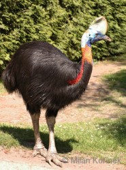
Kasuari Burung Paling Berbahaya Di Dunia
Kasuari merupakan sebangsa burung yang mempunyai ukuran tubuh sangat besar dan tidak mampu terbang. Kasuari yang merupakan binatang yang dilindungi di Indonesia dan juga menjadi fauna identitas provinsi Papua Barat terdiri atas tiga jenis (spesies). Ketiga spesies Kasuari yaitu Kasuari Gelambir Tunggal (Casuarius unappendiculatus), Kasuari Gelambir Ganda (Casuarius casuarius), dan Kasuari Kerdil (Casuarius bennetti).
Burung Kasuari merupakan burung besar yang indah menawan. Namun dibalik keindahan burung Kasuari mempunyai sifat yang agresif dan cenderung galak jika diganggu. Burung bergrnus Casuarius ini sangat galak dan pemarah dan tidak segan-segan mengejar ‘korban’ atau para pengganggunya. Karenanya di kebun binatangpun, Kasuari tidak dibiarkan berkeliaran bebas. Bahkan konon, The Guinnes Book of Records memasukkan burung Kasuari sebagai burung paling berbahaya di dunia. Meski untuk rekor ini saya belum dapat melakukan verifikasi ke situs The Guinness Book of Records.
Kasuari merupakan burung endemik yang hanya hidup di pulau Papua dan sekitarnya, kecuali Kasuari Gelambir Ganda (Casuarius casuarius) yang dapat juga ditemukan di benua Australia bagian timur laut. Dalam bahasa Inggris, Kasuari Gelambir Ganda (Casuarius casuarius) disebut (Southern Cassowary), Kasuari Gelambir Tunggal (Casuarius unappendiculatus) disebut (Northern Cassowary) dan Kasuari Kerdil (Casuarius bennetti) disebut sebagai (Dwarf Cassowary).
Ciri-ciri dan Tingkah Laku. Burung Kasuari mempunyai ukuran tubuh yang berukuran sangat besar, kecuali Kasuari Kerdil (Casuarius bennetti) yang ukuran tubuhnya lebih kecil. Burung Kasuari tidak dapat terbang. Burung kasuari dewasa mempunyai tinggi mencapai 170 cm, dan memiliki bulu berwarna hitam yang keras dan kaku.
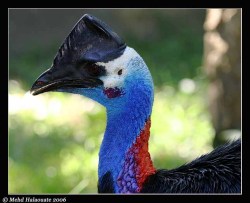
Di atas kepalanya Kasuari memiliki tanduk yang tinggi berwarna kecokelatan. Burung betina serupa dengan burung jantan, dan biasanya berukuran lebih besar dan lebih dominan.Kaki burung Kasuari sangat panjang dan kuat. Kaki ini menjadi senjata utama burung langka dan dilindungi ini. Kaki burung Kasuari mampu menendang dan merobohkan musuh-musuhnya, termasuk manusia, hanya dengan sekali tendangan. Mungkin karena tendangan dan agresifitasnya ini tidak berlebihan jika kemudian The Guinness Book of Records menganugerahinya sebagai burung paling berbahaya di dunia.
Pada Kasuari Gelambir Ganda terdapat dua buah gelambir berwarna merah pada lehernya dengan kulit leher berwarna biru.. Sedangkan pada Kasuari Gelambir Tunggal (Casuarius unappendiculatus), sesuai namanya hanya mempunyai satu gelambir.
Burung Kasuari yang termasuk satwa yang dilindungi dari keounahan ini memakan buah-buahan yang jatuh dari pohonnya. Burung Kasuari biasa hidup sendiri, dan berpasangan hanya pada saat musim kawin saja. Anak burung dierami oleh Kasuari jantan.
Meskipun Kasuari memiliki tubuh yang besar, namun ternyata tidak banyak yang diketahui tentang burung endemik papua ini. Apalagi untuk spesies Kasuari Gelambir Tunggal (Casuarius unappendiculatus) dan Kasuari Kerdil (Casuarius bennetti).Habitat dan Penyebaran. Burung Kasuari Gelambir Tunggal (Casuarius unappendiculatus) dan Kasuari Kerdil (Casuarius bennetti) merupakan satwa endemik pulau Papua (Indonesia dan Papua New Guinea), sedangkan Kasuari Gelambir Ganda (Casuarius casuarius) selain di pulau Papua juga terdapat di pulau Seram (Maluku, Indonesia) dan Australian bagian timur laut. Burung Kasuari mempunyai habitat di daerah hutan dataran rendah termasuk di daerah rawa-rawa.
Populasi dan Konservasi. Populasi burung Kasuari tidak diketahui dengan pasti namun diyakini dari hari ke hari semakin mengalami penurunan. Karena itu IUCN Redlist memasukkan burung Kasuari Gelambir Ganda (Casuarius casuarius) dan Kasuari Gelambir Tunggal (Casuarius unappendiculatus) dalam status konservasi Vulnerable (Rentan) sejak tahun 1994. Sedang Kasuari Kerdil (Casuarius bennetti) diberikan status konservasi Near Threatened (Hampir Terancam). Ancaman kepunahan burung Kasuari lebih karena perburuan baik untuk mendpatkan daging, bulu ataupun telurnya.
Klasifikasi ilmiah: Kerajaan: Animalia; Filum: Chordata; Kelas: Aves; Ordo: Struthioniformes; Famili: Casuariidae; Genus: Casuarius; Spesies: Casuarius casuarius, Casuarius unappendiculatus dan Casuarius bennetti.http://alamendah.wordpress.com/2010/04/12/kasuari-burung-paling-berbahaya-di-dunia/
Anggrek Hartinah Anggrek Tien Soeharto
Anggrek Hartinah atau Anggrek Tien Soeharto (Cymbidium hartinahianum) merupakan salah satu jenis tumbuhan anggrek yang endemik (hanya tumbuh di daerah tertentu) Sumater Utara, Indonesia. Jenis anggrek yang diketemukan pertama kali pada tahun 1976 ini bisa diketemukan di Desa Baniara Tele Kecamatan Harian Kabupaten Samosir, Sumatera Utara. Anggrek Tien Soeharto atau sering disebut juga sebagai Anggrek Hartinah (Cymbidium hartinahianum) merupakan anggrek tanah yang hidup merumpun.
Anggrek Tien Soeharto ini mengingatkan saya akan komentar Pakde Cholik, pemilik blog abdulcholik.com dalam postingan Alamendah Blog yang berjudul Perbedaan Rafflesia Arnoldii dan Bunga Bangkai. Terlontarlah sebuah pertanyaan; “…Apakah ada bunga atau tumbuhan yang namanya diambil dari nama orang Indonesia ?…”. Sebuah pertanyaan yang terlahir dari keresahan akan sederet penemuan satwa dan flora Indonesia yang banyak dilakukan oleh peneliti asing sedangkan peneliti bangsa sendiri terkesan lebih sukan di balik meja. Sehingga berbagai tanaman Indonesia pun mempunyai nama asing.
Di antara tumbuhan Indonesia yang dinamai dengan nama orang Indonesia adalah Anggrek Tien Soeharto atau disebut juga Anggrek Hartinah yang dalam bahasa latinnya disebut Cymbidium hartinahianum. Anggrek ini ini “>pertama kali ditemukan oleh Rusdi E Nasution, seorang peneliti dari Herbarium LBN/LIPI Bogor pada tahun 1976. Ketika itu anggrek ini tidak ditemukan dalam berbagai pusta maupun dalam koleksi. Kemudian oleh peneliti tersebut bersama peneliti lainnya J.B. Comber memberi nama ilmiah Cymbidium hartinahianum yang juga berarti anggrek Tien Soeharto pada hasil temuannya.
Penabalan (penamaan) pada jenis anggrek ini merupakan penghargaan atas jasa-jasa Ibu Tien Soeharto dalam rangka pengembangan dunia peranggrekan di Indonesia.
Ciri-ciri Fisik Anggrek Hartinah
Anggrek Hartinah (Tien Soeharto) merupakan salah satu anggrek tanah dengan pertumbuhan merumpun. Spesies anggrek ini menyukai tempat terbuka diantara rerumputan serta tanaman lain seperti jenis paku-pakuan, kantong semar, dan lain-lain pada ketinggian 1.700 meter diatas permukaan laut.
Daunnya berbentuk pita berujung meruncing dengan panjang 50-60 cm. Bunganya berbentuk bintang bertekstur tebal. Daun kelopak dan daun mahkotanya hampir sama besar, permukaan atasnya berwarna kuning kehijauan dan permukaan bawahnya kecoklatan dengan warna kuning pada bagian tepinya.
Konservasi Anggrek Hartinah
Anggrek Hartinah atau Tien Soeharto (Cymbidium hartinahianum) merupakan tumbuhan endemik Sumatera Utara sehingga dalam habitat alami hanya dapat diketemukan di Sumatera Utara saja. Habitatnya dapat ditemukan di Desa Baniara Tele Kecamatan Harian Kabupaten Samosir (berbatasan dengan Kabupaten Dairi).
Anggrek Hartinah ini bersama puluhan anggrek lainnya seperti anggrek Hitam, Anggrek Bulan Bintang dan lain-lain dikategorikan sebagai tanaman yang dilindungi berdasarkan Peraturan pemerintah Nomor 7 tahun 1999. Sehingga tumbuhan berfamili Orchidaceae ini tidak diperbolehkan diperjualbelikan kecuali untuk generasi ketiga. Generasi ketiga adalah tumbuhan hasil penangkaran yang telah mendapatkan izin dari pihak yang berwenang, biasanya BKSD.
Anggrek Hartinah ini telah dapat ditangkarkan di luar habitat aslinya. Salah satunya adalah di Kebun Raya Bogor.
Klasifikasi ilmiah: Kingdom: Plantae (Tumbuhan); Subkingdom: Tracheobionta (Tumbuhan berpembuluh); Super Divisi: Spermatophyta (Menghasilkan biji); Divisi: Magnoliophyta (Tumbuhan berbunga); Kelas: Liliopsida (berkeping satu / monokotil); Sub Kelas: Liliidae; Ordo: Orchidales; Famili: Orchidaceae (anggrek-anggrekan); Genus: Cymbidium; Spesies: Cymbidium hartinahianum
http://alamendah.wordpress.com/2009/09/13/anggrek-hartinah-anggrek-tien-soeharto/
Anggrek Bulan Puspa Pesona Indonesia
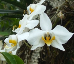
Anggrek bulan (Phalaenopsis amabilis) merupakan salah satu bunga nasional Indonesia, Anggrek bulan (Phalaenopsis amabilis) ditetapkan sebagai Puspa Pesona Indonesia mendampingi bunga melati (Jasminum sambac) yang ditetapkan sebagai puspa bangsa Indonesia dan padma raksasa (Rafflesia arnoldii) sebagai puspa langka Indonesia.
Anggrek bulan (Phalaenopsis amabilis) merupakan salah satu anggota genus Phalaenopsis, genus yang pertama kali ditemukan oleh seorang ahli botani Belanda, Dr. C.L. Blume. Phalaenopsis sendiri sedikitnya terdiri atas 60 jenis (spesies) dengan sekitar 140 varietas yang 60 varietas diantaranya terdapat di Indonesia.
Di Indonesia, anggrek bulan (Phalaenopsis amabilis) pertama kali ditemukan di Maluku. Anggrek bulan memiliki beberapa nama daerah seperti anggrek wulan (Jawa dan Bali), anggrek terbang (Maluku), dan anggrek menur (Jawa). Pemerintah menetapkan anggrek bulan sebagai puspa pesona mendampingi melati (puspa bangsa), dan padma raksasa (puspa langka) berdasarkan Keputusan Presiden Republik Indonesia Nomor 4 Tahun 1993.
Pesona Anggrek Bulan. Anggrek bulan (Phalaenopsis amabilis) merupakan jenis anggrek (Orchidaceae) yang mempunyai ciri khas kelopak bunga yang lebar dan berwarna putih. Meskipun saat ini sudah banyak anggrek bulan hasil persilangan (anggrek bulan hibrida) yang memiliki corak dan warna beragam jenis.
Anggrek bulan (Phalaenopsis amabilis) termasuk dalam tanaman anggrek monopodial yang menyukai sedikit cahaya matahari sebagai penunjang hidupnya. Daunnya berwarna hijau dengan bentuk memanjang. Akar anggrek bulan berwarna putih berbentuk bulat memanjang dan terasa berdaging. Bunga anggrek bulan memiliki sedikit keharuman dan waktu mekar yang lama serta dapat tumbuh hingga diameter 10 cm lebih.
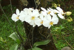 Anggrek bulan (Phalaenopsis amabilis) tumbuh liar dan tersebar luas mulai dari Malaysia, Indonesia, Filipina, Papua, hingga ke Australia. Anggrek bulan hidup secara epifit dengan menempel pada batang atau cabang pohon di hutan-hutan. Secara liar anggrek bulan mampu tumbuh subur hingga ketinggian 600 meter dpl.
Lantaran keindahannya itu wajar jika kemudian anggrek bulan ditetapkan sebagai puspa pesona, satu diantara 3 bunga nasional Indonesia. Anggrek bulan ditetapkan sebagai puspa pesona mendampingi melati (puspa bangsa) dan padma raksasa (puspa langka).
Meskipun banyak pehobi anggrek yang membudidayakan anggrek bulan. Juga banyak yang melakukan persilangan sehingga memunculkan varietas-varietas baru anggrek bulan hibrida, namun kelestarian puspa pesona ini di alam liar tetap semakin terdesak oleh hilangnya habitat sebagai akibat deforestasi hutan baik akibat penebangan liar ataupun kebakaran hutan.
Anggrek bulan di alam liar kini membutuhkan perhatian tersendiri. Jangan sampai sang puspa pesona memudar pesonanya.
Klasifikasi ilmiah. Kerajaan: Plantae; Ordo: Asparagales; Familia: Orchidaceae; Subsuku: Epidendroideae; Genus: Phalaenopsis; Spesies: Phalaenopsis amabilis
Sinonim: Epidendrum amabile L. (basionym); Cymbidium amabile (L.) Roxb.; Synadena amabilis (L.) Raf.; Phalaenopsis grandiflora Lindl.; Phalaenopsis grandiflora var. aurea auct.; Phalaenopsis amabilis var. aurea (auct.) Rolfe; Phalaenopsis gloriosa Rchb.f.
http://alamendah.wordpress.com/2010/04/23/anggrek-bulan-puspa-pesona-indonesia/
Mengenal Bunga Sedap Malam (Polianthes tuberosa)
Bunga Sedap Malam atau Polianthes tuberosa adalah nama salah satu bunga yang banyak sudah dikenal luas di Indonesia sebagai bunga potong dan penghasil parfum. Bunga Sedap Malam juga telah ditetapkan sebagai flora identitas provinsi Jawa Timur mendampingi Bekisar sebagai Fauna Identitas Provinsinya.
Bunga Sedap Malam sebenarnya bukan bunga asli Indonesia. Diperkirakan bunga ini berasal dari Meksiko dan telah diintroduksi ke Indonesia sejak masuknya bangsa Eropa dan China ke Indonesia.
Disebut sebagai Bunga Sedap Malam lantaran bunga ini biasa mekar dan menebar aroma wangi pada malam hari. Selain disebut Sedap Malam, di Melayu bunga ini dikenal juga sebagai Sundal Malam. Tanaman ini dalam bahasa Inggris dikenal sebagai Tuberose. Sedangkan dalam bahasa latin tanaman ini dinamai Polianthes tuberosa. 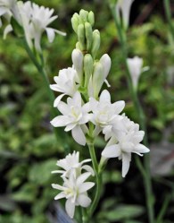

Ciri dan Diskripsi. Bunga Sedap Malam tumbuh merumpun dengan tinggi sekitar 0,5 – 1,5 meter. Serumpun batangnya tumbuh dari satu atau beberapa umbi induk dan beberapa umbi anak. Umbi ini merupakan batang semu sekaligus sebagai penyimpan makanan. Umbi bunga Sedap Malam juga digunakan untuk perbanyakan tanaman secara vegetatif.
Daun bunga Sedap Malam (Polianthes tuberosa) berbentuk panjang pipih berwarna hijau mengkilat di bagian permukaan atas dan hijau muda pada bagian permukaan bawah daun. Pada pangkal daun terdapat bintik-bintik berwarna kemerah-merahan. Daun dapat berukuran hingga sepanjang 60 cm.
Tangkai bunga muncul di ujung tanaman berbentuk memanjang dan beruas-ruas. Di setiap ruas muncul daun bunga yang berbentuk pipih memanjang dengan ukuran lebih kecil dari daun biasa. Pada tangkai bunga melekat 5-12 kuntum bunga (terkadang lebih) dengan mahkota bunga berwarna putih dan sedikit kemerahan di bagian ujung.
Mekarnya bunga Sedap Malam (Polianthes tuberosa) tidak serempak melainkan berurutan. Kuntum bunga bagian bawah akan mekar terlebih dahulu lalu menyusul kuntum-kumtum bunga di atasnya secara berurutan.
Bunga Sedap Malam dikenal memiliki kesegaran yang mampu bertahan lama. Meskipun telah dipotong bunga yang menjadi flora Identitas provinsi Jawa Timur ini kesegarannya dapat bertahan selama 5-10 hari.
Pemanfaatan. Bunga Sedap Malam (Polianthes tuberosa) banyak dibudidayakan di berbagai daerah di Indonesia. Bunga ini banyak dimanfaatkan sebagai bunga potong untuk berbagai keperluan. Selain itu bunga Sedap Malam juga dapat diolah sebagai bahan pembuat parfum.
Klasifikasi ilmiah: Kerajaan: Plantae; Divisi: Magnoliophyta; Kelas: Liliopsida; Ordo: Asparagales; Famili: Agavaceae; Genus: Polianthes; Spesies: Polianthes tuberosa.
http://alamendah.wordpress.com/2011/03/27/mengenal-bunga-sedap-malam-polianthes-tuberosa/
Anggrek Larat (Dendrobium phalaenopsis) Anggrek Langka dari Maluku
Anggrek Larat (Dendrobium phalaenopsis) termasuk anggrek langka dari Maluku. Bahkan anggrek Larat termasuk satu dari 12 spesies anggrek langka yang dilindungi di Indonesia. Anggrek Larat (Dendrobium phalaenopsis) juga ditetapkan sebagai flora identitas provinsi Maluku.
Anggrek ini dinamakan Anggrek Larat lantaran pertama kali ditemukan di pulau Larat, Tanimbar, Maluku. Namun lantaran keindahannya itu, semakin hari anggrek larat semakin langka di habitat aslinya.
Anggrek Larat yang dalam bahasa Inggris dikenal sebagai Cooktown Orchid, berkerabat dekat dengan beberapa jenis anggrek lainnya seperti Anggrek Merpati, Anggrek Albert, Anggrek Stuberi, Anggrek Jamrud, Anggrek Karawai, dan Anggrek Kelembai. Dalam bahasa latin tumbuhan ini dikenal sebagai Dendrobium phalaenopsis dengan sinonim Vappodes phalaenopsis, dan Dendrobium bigibbum.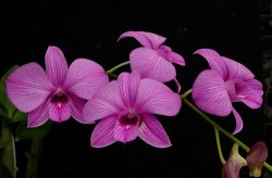 Diskripsi Anggrek Larat. Anggrek Larat yang ditetapkan sebagai flora identitas provinsi Maluku ini mempunyai batang berbentuk gada dengan pangkal berukuran kecil, bagian tengah membesar dan ujungnya mengecil kembali. Daun Anggrek Larat (Dendrobium phalaenopsis) berbentuk lanset dengan ujung tidak simetris. Panjang daunnya kira-kira 12 cm, dengan lebar kira-kira 2 cm.
 Diskripsi Anggrek Larat. Anggrek Larat yang ditetapkan sebagai flora identitas provinsi Maluku ini mempunyai batang berbentuk gada dengan pangkal berukuran kecil, bagian tengah membesar dan ujungnya mengecil kembali. Daun Anggrek Larat (Dendrobium phalaenopsis) berbentuk lanset dengan ujung tidak simetris. Panjang daunnya kira-kira 12 cm, dengan lebar kira-kira 2 cm.
Bunga Anggrek Larat berwarna keungunan pucat hingga ungu tua. Tersusun dalam bentuk tandan yang tumbuh pada buku-buku batangnya, agak menggantung. Panjang tandan bunganya kurang lebih 60 cm dengan jumlah bunga tiap tandan 6 – 24 kuntum. Masing-masing bunga bergaris tengah kurang lebih 6 cm. Daun Kelopak berbentuk lanset, berwarna keunguan. Daun Mahkota lebih pendek, tetapi lebih lebar dari pada kelopaknya. Pangkalnya sempit dengan ujungnya runcing dan berwarna keunguan. Bibir bertajuk tiga membentuk corong dengan tajuk tengahnya yang lebar, runcing atau meruncing. Buah berbentuk jorong, panjang 3,2 cm namun bunganya jarang menjadi buah. 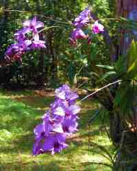

Anggrek Larat (Dendrobium phalaenopsis) yang pertama kali di temukan di pulau Larat, Maluku tumbuh baik di daerah panas, pada ketinggian antara 0 – 150 m dpl. Di habitat aslinya, Anggrek yang dijadikan bunga maskot provinsi Maluku ini tumbuh pada pohon-pohonan dan karang-karangan kapur yang mendapat sinar matahari cukup.
Konservasi Anggrek Larat. Anggrek Larat pernah menjadi sangat terkenal di kalangan para pecinta Anggrek, di samping Anggrek Bulan (Phalaenopsis amabilis). Karenanya hingga saat ini banyak sekali anggrek hibrida komersial dendrobium yang merupakan hasil persilangan dari anggrek spesies (anggrek alami) jenis ini. 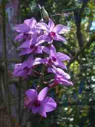

Mungkin lantaran itu, di habitat aslinya anggrek Larat semakin langka dan terancam punah. Bunga anggrek yang kemudian ditetapkan sebagai flora identitas provinsi Maluku ini akhirnya ditetapkan menjadfi salah satu dari 12 spesies Anggrek yang langka dan dilindungi di Indonesia berdasarkan Peraturan Pemerintah No. 7 Tahun 1999.
Semoga Si Ungu dari pulau Larat ini masih berkesempatan untuk menebarkan pesona keindahanya di habitat aslinya.
Klasifikasi Ilmiah. Kerajaan: Plantae; Divisi: Magnoliophyta; Kelas: Liliopsida; Ordo: Orchidales; Famili: Orchidaceae; Genus: Dendrobium; Spesies: Dendrobium phalaenopsis.
http://alamendah.wordpress.com/2011/03/09/anggrek-larat-dendrobium-phalaenopsis-anggrek-langka-dari-maluku/
Teratai Tanaman Air dengan Bunga Mempesona
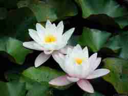
Teratai merupakan nama umum untuk genus Nymphaea yang merupakan tumbuhan air. Tanaman teratai memiliki ciri khas dengan daun yang mengambang di permukaan air yang tenang. Tanaman teratai pun menghasilkan bunga mempesona yang memiliki warna beraneka ragam.
Di beberapa daerah di Indonesia teratai dikenal dengan beberapa nama yang hampir mirip seperti teratai, dan terate. Dalam bahasa Inggris, bunga dari genus Nymphaea ini dikenal sebagai water-lily atau waterlily.
Terdapat lebih dari 50 jenis (spesies) teratai di dunia yang tersebar mulai dari daerah tropis hingga subtropis. Konon spesies-spesies teratai tropis berasal dari Mesir.
Ciri-ciri. Tanaman teratai tumbuh di permukaan air yang tenang. Tanaman teratai memiliki daun yang tumbuh mengambang di permukaan air. Bunga teratai juga terdapat di permukaan air, bunga dan daun teratai keluar dari tangkai yang berasal dari rizoma yang berada di dalam lumpur pada dasar kolam, sungai atau rawa.
Tangkai teratai terdapat di tengah-tengah daun. Daun berbentuk bundar atau bentuk oval yang lebar yang terpotong pada jari-jari menuju ke tangkai. Permukaan daun tidak mengandung lapisan lilin sehingga air yang jatuh ke permukaan daun tidak membentuk butiran air.
Bunga teratai tumbuh pada tangkai yang merupakan perpanjangan dari rimpang. Diameter bunga bergenus Nymphaea
ini antara 5-10 cm.
ini antara 5-10 cm.
Ragam Jenis Spesies. Beberapa macam jenis tanaman teratai antara lain:
- Nymphaea alba – European White Water-lily (teratai putih)
- Nymphaea amazonum
- Nymphaea ampla
- Nymphaea blanda
- Nymphaea caerulea – Egyptian Blue Water-lily
- Nymphaea calliantha
- Nymphaea candida
- Nymphaea capensis – Cape Blue Water-lily
- Nymphaea citrina
- Nymphaea colorata
- Nymphaea elegans
- Nymphaea fennica
- Nymphaea flavovirens
- Nymphaea gardneriana
- Nymphaea gigantea – Australian Water-lily
- Nymphaea heudelotii
- Nymphaea jamesoniana
- Nymphaea leibergii – Dwarf Water-lily
- Nymphaea lotus – Egyptian White Water-lily (Teratai kecil)
- Nymphaea lotus var. termalis
- Nymphaeae lutea
- Nymphaea macrosperma – Native to Australia’s Top End
- Nymphaea mexicana – Yellow Water-lily
- Nymphaea micrantha
- Nymphaea nouchali – Red and blue water lily (Bunga Nasional Sri Lanka)
- Nymphaea odorata – Fragrant Water-lily
- Nymphaea pubescens – Hairy water lily (Bunga Nasional Bangladesh)
- Nymphaea rubra – India Red Water-lily (Teratai merah)
- Nymphaea rudgeana
- Nymphaea stellata
- Nymphaea stuhlmannii
- Nymphaea sulfurea
- Nymphaea tetragona – Pygmy Water-lily (Teratai kerdil)
- Nymphaea thermarum
Manfaat. Tanaman teratai banyak dimanfaatkan sebagai tanaman hias. Namun selain sebagai tanaman hias, teratai juga memiliki khasiat sebagai tanaman obat-obatan tradisional yang antara lain dapat mengobati penyakit, diare, disentri, keputihan, kanker nasopharynx, demam, insomnia; Hipertensi, muntah darah, mimisan, batuk darah, sakit jantung; Beri-beri, sakit kepala, berak dan kencing darah, anemia, ejakulasi dini.
Teratai Bukan Seroja. Sebagian orang masih sering mencampuradukkan dan menganggap sama antara teratai (genus Nymphaea) dan seroja (genus Nelumbo). Bahkan dulu dianggap berkerabat dekat. Nyatanya keduanya sangat berbeda. Bunga seroja berbeda dengan teratai. Pada Nelumbo, bunga terdapat di atas permukaan air (tidak mengapung), kelopak bersemu merah (teratai berwarna putih hingga kuning), daun berbentuk lingkaran penuh dan rimpangnya biasa dikonsumsi.
Klasifikasi ilmiah: Kerajaan: Plantae; Subkingdom: Tracheobionta (Tumbuhan berpembuluh); Super Divisi: Spermatophyta (Menghasilkan biji); Divisi: Magnoliophyta (Tumbuhan berbunga); Kelas: Magnoliopsida (berkeping dua / dikotil); Sub Kelas: Magnoliidae; Ordo: Nymphaeales; Famili: Nymphaeaceae; Genus: Nymphaea; Spesies: lihat artikel.
10 Misteri Otak Manusia yang Belum Terpecahkan
10 Misteri Otak Manusia yang Belum Terpecahkan

Pasti bertanya – tanya deh, maksud “Misteri Otak Manusia” itu gimana ? haha , baca deh , ni juga dapet pas lagi browsing pake keyword “Otak”.
Pendahuluan
Hmmm , Jangankan alam sekitar, diri sendiri kita pun masih banyak menyimpan tanda tanya. Otak manusia bisa disamakan dengan prosesor komputer. Bedanya, kinerja prosesor dapat diuraikan secara logika, sedangkan otak kita tidak.
Ada 10 misteri yang masih menyelubungi seluk beluk otak manusia. Ilmuwan masih terus mencoba mencari penjelasan ilmiahnya. Tapi tetap saja misteri itu merupakan rahasia kehidupan ciptaan Tuhan yang luar biasa.
Berikut 10 misteri seputar otak manusia yang kita alami sehari-hari, tapi tetap kita tak mampu mencari penyebabnya.
1. Kesadaran
Saat bangun di pagi hari, kita tersadar dari tidur. Menikmati sinar matahari dari celah jendela, udara pagi nan sejuk, dan seterusnya. Kita menyebutnya sebagai kesadaran. Bidang ini memicu topik majemuk yang dibahas ilmuwan sejak zaman dulu. Pakar neurologi mutakhir menjabarkan kesadaran sebagai suatu topik riset realistis.
2. Hidup Membeku
Hidup abadi memang hanya ada dalam khayalan manusia. Namun ilmuwan telah menemukan cryonic, temuan yang mampu membuat manusia memiliki dua kehidupan. Salah satu pusat cryonic adalah Alcor Life Extension Foundation, di Arizona, yang menyimpan tubuh mahluk hidup dalam tabung berisi nitrogen cair dengan suhu minus 320 fahrenheit.
Idenya adalah manusia yang sudah meninggal akibat penyakit akan dicairkan dan dihidupkan kembali di masa mendatang saat penyakit itu sudah bisa disembuhkan.
Jenazah Ted Williams, pemain baseball kenamaan disimpan di sini. Karena teknologinya belum ditemukan, maka penghidupan kembali belum dilakukan. namun tubuhnya sudah “dilelehkan” dengan suhu yang tepat sehingga sel-selnya membeku dan memecah.
3. Misteri Kematian
Bagaimana manusia menjadi tua ? manusia terlahir dengan mekanisme tubuh yang mampu bertahan dari penyakit. Itu sebabnya luka bisa sembuh sendiri tanpa diobati. Tapi seiring dengan bertambah usia, mekanisme itu menurun.
Kenapa bisa begitu ? Ada dua teori penjelasannya.
· Pertama, penuaan adalah bagian dari genetika manusia.
· Kedua, penuaan adalah hasil dari sel-sel tubuh yang rusak.
4. Alam VS Asuhan
Perdebatan tentang pikiran dan kepribadian manusia masih berkutat antara dua hal di atas. Kepribadian dan pemikiran manusia dikatakan dikontrol oleh gen atau lingkungan ? Atau bisa jadi keduanya ? Masih belum ada kesepakatan di kalangan ilmuwan tentang hal ini.
5. Pemicu Otak
Tertawa adalah hal yang paling sedikit dipahami dari perilaku manusia. Para ilmuwan menemukan bahwa selama tertawa, ada tiga bagian otak yang terlibat.
· Pertama, bagian yang berpikir sebelum kita memahami suatu gurauan.
· Kedua, area yang bergerak untuk memberitahu otot kita untuk melakukan sesuatu.
· Lalu sebuah area emosional yang menggugah perasaan geli.
John Morreall, ilmuwan peneliti humor dari College of William and Mary, menemukan bahwa tertawa adalah respon bermain atas kisah yang tidak sesuai dengan harapan. Tertawa juga mampu menular pada orang lain.
6. Daya Ingat
Beberapa pengalaman sulit dilupakan, sebaliknya kita justru kerap melupakan hal-hal penting. Bagaimana itu bisa terjadi ?
Menggunakan teknik pencitraan otak, ilmuwan menemukan adanya mekanisme yang bertanggungjawab pada penciptaan dan penyimpanan memori. Mereka menemukan hippocampus dan materi abu-abu otak yang berperan sebagai kotak memori. Tapi mengapa ada memori yang mudah diingat dan dilupakan, masih tetap jadi misteri.
7. Jam Biologis
Otak juga memiliki nukleus suprachiasmatic nucleus alias jam biologi. Bagian ini memprogram tubuh untuk mengikuti irama waktu 24 jam. Jam biologi juga menyesuaikan suhu tubuh, siklus bangun tidur, juga produksi hormon melatonin. Perdebatan terakhir adalah apakah suplemen melatonin mampu mencegah jet lag ?
8. Perasaan Dihantui
Diperkirakan 80 persen dari sensasi pengalaman termasuk gatal, tertekan, nyaman dan rasa sakit datang dari bagian tubuh yang hilang. Ada orang yang mengalami adanya organ tubuh mereka yang tidak nampak tapi bisa merasakan.
Salah satu penjelasan adalah adanya area syaraf di salah satu organ tubuh yang menciptakan konseksi baru pada saraf tulang belakang dan berlanjut mengirimkan sinyal ke otak.
9. Tidur
Mengapa manusia butuh tidur ? Ilmuwan paham bahwa semua mamalia butuh tidur cukup. Tidak cukup tidur berkepanjangan akan menimbulkan halunisasi bahkan kematian.
Ada dua tingkatan dalam tidur, yakni :
· tidur yang non-rapid eye movement (NREM), terjadi selama otak memperlihatkan rendahnya aktivitas metabolik.
· Lalu tidur tingkat rapid eye movement (REM), saat otak masih cukup aktif.
10. Mimpi
Selain tidur, mimpi juga menjadi misteri. Kemungkinannya adalah, bermimpi merupakan latihan otak yang menstimulasi trafik synap antar sel-sel otak. Teori lain mengatakan manusia bermimpi mengenai tugas dan emosinya yang tak sempat diperhatikan selama mereka terjaga di siang hari.
http://gozarago.blogspot.com/2010/06/10-misteri-otak-manusia-yang-belum.html
Langganan:
Postingan (Atom)


